

Feeling a Dent in My Head: What You Need to Know
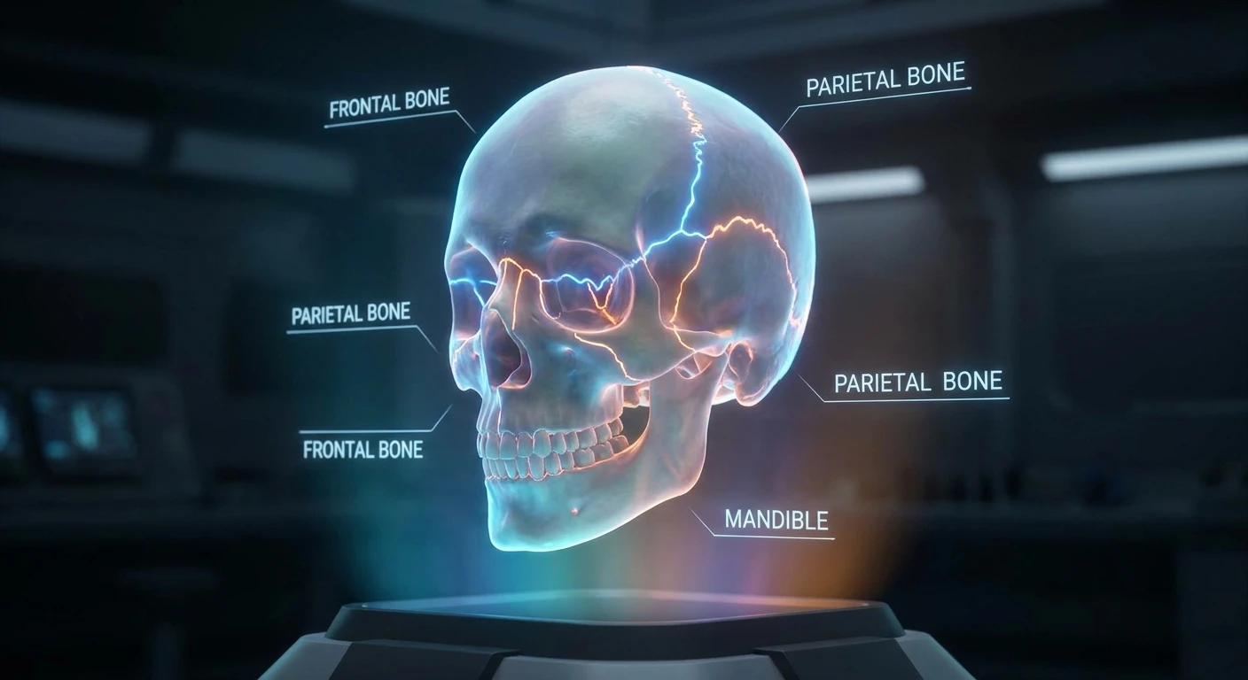
When you suddenly notice a dent in my head, panic is often the first reaction that floods your mind. You might be running your fingers through your hair, washing it in the shower, or just resting your hand on your scalp while reading a book when you feel an unexpected, unfamiliar dip. The human skull is far from a perfectly smooth, symmetrical bowling ball. Instead, it is a highly complex biological structure made up of multiple interlocking bones that fuse together during infancy and childhood. Those natural fusion lines, known medically as cranial sutures, frequently leave slight ridges, bumps, or depressions that are completely natural and almost always harmless. Understanding the structural layout of your cranium helps immediately ease the fear that something is terribly wrong. Most people have minor irregularities on the top, back, or sides of their skulls without ever realizing it until they happen to touch that exact spot. However, certain changes in the shape of your head can sometimes point to underlying medical conditions like previous trauma, bone metabolic diseases, or benign cysts. By closely examining the biological makeup of the cranium, the historical understandings of skull shapes, the scientific biomechanics of our bone structures, and establishing a clear step-by-step plan for assessing any unusual cranial contours, you can take control of your health. Getting familiar with your own anatomy provides invaluable peace of mind and equips you with the right knowledge and vocabulary for your next medical checkup.
Core Reasons Behind Skull Irregularities
Discovering a structural groove or dip in your scalp requires a logical, calm examination of what actually sits beneath the skin. The primary reason for a newly discovered indentation usually boils down to the natural, baseline anatomy of the human skull, which consists of twenty-two different bones protecting the brain and forming the face. Where these bones meet, especially along the sagittal suture at the top of the head or the coronal suture near the front, you can frequently feel a slight depression, ridge, or groove. It is entirely normal to feel these features, particularly if you have recently changed your hairstyle, shaved your head, lost a significant amount of weight, or simply never paid close attention to that specific anatomical spot before.
Read also: Pain Under Armpit: Causes, Symptoms, and Complete Action Pla; Ear Drainage Causes and Quick Treatments.
Beyond natural anatomy, several external and internal factors can also create tangible indentations. Minor trauma from a childhood injury might have healed with a slight permanent depression in the outer bone layer. More serious conditions, such as Gorham’s disease, Paget’s disease of bone, or even certain types of benign subcutaneous cysts, can alter the bone’s outer layer or the tissue sitting immediately above it.
Here is a detailed breakdown of common cranial irregularities, their typical severity, and accompanying symptoms:
| Cause of Depression | Severity Level | Typical Associated Symptoms |
|---|---|---|
| Natural Cranial Sutures | Harmless | None; completely painless, static, and present since early childhood. |
| Healed Depressed Fracture | Moderate (Requires past medical context) | Previous history of head trauma, occasional localized tenderness, static size. |
| Subcutaneous Cysts or Lipomas | Mild to Moderate | Soft or rubbery feel, slowly changing shape over time, easily movable under the skin. |
| Paget’s Disease of Bone | Severe (Requires medical intervention) | Bone pain, localized warmth, hearing loss, visible enlargement of the skull. |
If you are assessing the situation at home, here are a few practical examples of what to evaluate:
- A natural sagittal suture will run directly down the middle of the top of your head, feeling like a slight valley between two gentle ridges, running from front to back.
- A healed injury from a childhood sports accident will remain unchanged in size over decades and typically has no pain associated with it in adulthood.
- A cyst might feel like a dent if the surrounding tissue is highly swollen, making the normal bone underneath feel artificially recessed by comparison.
Keep these three key evaluation points in mind when checking your scalp:
- Monitor the spot carefully for any rapid changes in overall depth, width, or shape.
- Note any accompanying physical sensations, such as chronic headaches, unexplainable dizziness, or sharp localized pain.
- Check if the skin over the depression is discolored, red, inflamed, or excessively flaky, which could indicate a dermatological issue rather than a bone problem.
Historical Perspectives on Skull Shapes
Origins of Cranial Mapping
The intense fascination with the shape, bumps, and dents of the human head is certainly not a new phenomenon. In the late eighteenth and early nineteenth centuries, a pseudo-science known as phrenology took the medical and psychological worlds by storm. Founded by the German physician Franz Joseph Gall, phrenology proposed that the contours of the skull directly corresponded to a person’s specific character traits, overall intelligence, and even their criminal tendencies. Practitioners of this flawed discipline would carefully run their hands over a patient’s head, feeling for every single ridge, bump, or dent. They mistakenly believed that a depression in a specific area meant a distinct lack of a certain personality trait, while a bump indicated an abundance of it. While completely debunked by modern science today, this early obsession inadvertently laid the historical groundwork for mapping the brain, sparking the initial idea that different physical parts of the brain control different neurological functions.
Evolution of Skull Anatomy Understanding
As modern medicine rapidly advanced through the nineteenth and twentieth centuries, competent doctors completely discarded the unfounded myths of phrenology and began studying the actual, verifiable osteology of the cranium. The groundbreaking invention of the X-ray by Wilhelm Conrad Röntgen in 1895 absolutely revolutionized how humanity viewed the skeletal system. Suddenly, physicians could literally look past the skin, muscle, and tissue to see the exact structure of the human skull without performing dangerous surgery. They quickly mapped out the fontanelles, the soft spots in an infant’s skull, and studied exactly how these biological gaps close and fuse, noting that they sometimes leave a small, permanent dent as a remnant of the process. This critical evolution of anatomical science shifted the global focus from guessing personality traits based on head shape to treating actual physical trauma, congenital anomalies, and bone diseases.
Modern State of Cranial Diagnostics
Today, if you visit a qualified neurologist, an osteopathic doctor, or an orthopedic specialist about an unusual skull depression, they rely on highly advanced imaging technology rather than just traditional manual palpation. High-resolution Computed Tomography (CT) scans and Magnetic Resonance Imaging (MRI) machines provide stunning, three-dimensional, cross-sectional images of the entire skull and the underlying brain matter. These incredible diagnostic tools allow medical professionals to differentiate instantly between a harmless natural suture, a benign fatty cyst, or a much more serious bone degradation issue. The modern state of cranial diagnostics ensures that any structural anomaly is evaluated with absolute pinpoint accuracy, providing patients with immediate reassurance or a clear, targeted, and effective treatment plan.
The Science Beneath the Scalp
The Biomechanics of the Cranium
Your skull is an absolute marvel of natural biological engineering, specifically designed to protect the most vital and delicate organ in your body. It is structurally composed of the neurocranium, which creates the protective vault surrounding the brain, and the viscerocranium, which forms the underlying structure of the face. The neurocranium is made of incredibly strong, flat bones connected by tough, fibrous joints called sutures. These sutures are not perfectly flat lines; they zig-zag heavily and interlock tightly like pieces of a complex jigsaw puzzle. This unique interlocking mechanism provides incredible structural strength and necessary flexibility, allowing the skull to absorb the shock of minor everyday impacts safely. The bone itself consists of three layers: a hard outer table, a hard inner table, and a spongy middle layer called the diploë. When you run your hand over your head and feel a subtle dip, you are often just feeling the natural meeting point of the frontal and parietal bones at the coronal suture, where the outer table dips slightly. Because these joints are essentially biological seams, they rarely lay perfectly flat against each other.
How Bone Remodeling Works
Bone is fundamentally not a static, unchanging material; rather, it is highly active, living, dynamic tissue that constantly breaks itself down and rebuilds itself throughout your entire life. This continuous process, known scientifically as bone remodeling, involves specialized cells called osteoclasts that actively break down older bone tissue, and osteoblasts that subsequently build strong new bone in its place. Sometimes, this remodeling process can be heavily influenced by prolonged external pressure, systemic health conditions, nutritional deficits, or normal aging, leading to slight changes in the surface texture and thickness of the skull over decades.
Consider these highly fascinating scientific facts about your cranium:
- The adult human skull is made up of 22 individual bones, excluding the tiny ossicles located deep in the middle ear.
- At the time of birth, a baby’s skull actually consists of 44 separate bony elements that slowly fuse together as the child grows and develops.
- The sagittal suture, which runs directly down the midline of the skull from front to back, typically does not completely fuse into solid bone until a person reaches their late thirties or early forties.
- Extensive osteology studies show that slight cranial asymmetry is biologically present in over 90 percent of the global human population, meaning practically no one’s head is perfectly round or smooth.
- Certain systemic conditions like severe vitamin D deficiency during early childhood, known as rickets, can permanently alter the structural density, thickness, and overall shape of the cranial vault for life.
- The spongy diploë layer between the hard skull plates contains red bone marrow, which is actively responsible for producing vital red blood cells.
Your 7-Step Action Plan for Cranial Anomalies
If you are currently fixated on a new, unexplained spot on your head, follow this practical, logical, seven-step assessment plan to evaluate the situation effectively.
Step 1: Do Not Panic and Stop Probing
The vast majority of newly discovered skull dents are completely benign anatomical features that have likely been there for years. The most crucial first step is to stop constantly pressing your fingers into it. Excessive probing can quickly irritate the delicate scalp tissue, cause localized swelling, and make the depression feel much larger and more painful than it actually is. Give the area a rest.
Step 2: Wash Your Hair and Inspect Visually
Hop in the shower and wash your hair thoroughly with normal shampoo. Wet hair lays perfectly flat against the scalp, allowing you to get a much clearer visual view of the skin in the mirror. Use two mirrors to inspect the area from multiple angles. Look closely for redness, dark moles, insect bites, or skin irregularities that might better explain the abnormal texture.
Step 3: Check for Associated Neurological Symptoms
Sit down quietly and do a quick, honest body scan. Are you currently experiencing unusual, severe headaches? Is your vision inexplicably blurry? Do you feel dizzy, nauseous, or is the dent extremely tender to the lightest touch? Write down any and all symptoms you are feeling right now, as this information is vital for a doctor.
Step 4: Photograph and Document the Area
Have a trusted friend or family member take a clear, well-lit, high-resolution photo of the area with their smartphone. Gently measure the approximate width and depth with a soft tape measure. Documenting the exact size gives you an objective baseline to determine if the spot is actually changing over time or if your anxiety is just making it seem larger.
Step 5: Consult a General Practitioner
If the dent appears entirely new, is actively painful, or is accompanied by any of the neurological symptoms mentioned in step three, schedule an appointment with your primary care doctor immediately. Bring your detailed documentation and explain clearly when you first noticed the physical change.
Step 6: Get Diagnostic Imaging if Recommended
Your doctor may prudently order a standard X-ray or a more detailed CT scan just to be absolutely safe. Always follow through with these diagnostic tests. They are completely quick, entirely painless, and provide definitive, unquestionable answers about what is happening beneath the layers of the skin.
Step 7: Follow Up and Monitor Calmly
If the doctor reviews the imaging and gives you a confident all-clear, simply monitor the area passively once a month. Keep a brief written log of any future changes. Trust the advanced medical imaging, accept the professional diagnosis, and intentionally let go of the anxiety once a medical professional has confirmed it is just a natural feature of your unique skull.
Myths and Realities About Skull Shapes
There is an incredible amount of misinformation floating around the internet regarding what different head shapes actually mean. We can clear up the confusion with hard medical facts.
Myth: Feeling a dent on your scalp means your brain is totally unprotected in that specific area.
Reality: The skull bones are incredibly thick and resilient. A slight surface depression in the outer bone table does not mean the bone is dangerously thin or that your brain is at any elevated risk from normal everyday activities.
Myth: Your skull stops changing shape completely after childhood ends.
Reality: While major structural growth definitely stops, biological bone remodeling continues actively throughout your entire life. Advanced age, shifting nutrition, and certain metabolic health conditions can slightly alter overall bone density and texture even well into your senior years.
Myth: A newly discovered dent is always a definitive sign of a brain tumor.
Reality: The vast majority of brain tumors grow deep inside the skull cavity and rarely cause inward dents on the tough outer bone. Most external dents are strictly related to the bone tissue itself, the overlying scalp tissue, or natural structural sutures, not the actual brain tissue underneath.
Myth: Wearing tight hats frequently can permanently dent your skull over time.
Reality: Hats can temporarily compress the soft skin, fat, and hair on your scalp, leaving a temporary red mark or groove, but they absolutely do not possess the force required to permanently reshape your solid, calcified skull bones.
Frequently Asked Questions & Conclusion
Can wearing heavy headphones cause a permanent dent?
No. Heavy gaming or studio headphones can temporarily compress the skin and fatty tissue on your scalp, creating a temporary groove, but they absolutely cannot permanently reshape your hard, calcified skull bones.
Are unique skull dents and ridges hereditary?
Yes, they often are. The general overall shape of your head and the specific prominence of your cranial sutures are largely dictated by your genetic lineage.
Does severe dehydration cause scalp dents?
In young infants, severe dehydration can indeed cause the fontanelle to sink noticeably. In healthy adults, however, the bones are fully fused, so dehydration does not cause hard bone depressions.
When should I immediately go to the ER for a head dent?
You must seek immediate emergency medical care if the dent appears immediately after severe head trauma, or if it is accompanied by vomiting, deep confusion, lethargy, or any loss of consciousness.
Can a local dermatologist help with scalp dents?
Yes, they frequently can. If the apparent dent is actually caused by a skin issue, a ruptured cyst, or a shifting lipoma, a board-certified dermatologist is the perfect medical specialist to diagnose and treat it.
Do adult humans still have soft spots on their heads?
No. The natural soft spots present at birth fully fuse and calcify during early childhood, turning into solid, durable bone sutures.
Can significant weight loss expose hidden skull dents?
Absolutely. Losing a significant amount of fat in your face and scalp reduces the padding over your bones, which can make natural bone ridges and natural dips much more prominent to the touch.
Can chronic stress or tension cause a dent?
While severe stress can cause tight scalp muscles, it cannot physically alter the shape of the cranial bone.
Are bone dents related to hair loss?
No. Male or female pattern baldness affects the hair follicles, not the underlying skeletal structure. However, losing hair makes existing bone contours much more visible.
Can sleeping on one side dent my head?
Adult skulls are far too dense to be reshaped by standard pillows or sleeping positions. This is only a concern for newborns whose skulls are highly malleable.
If you have recently discovered a dent in my head, always remember that human anatomy is uniquely and wonderfully textured. While it is easy to jump to conclusions, most cranial irregularities are nothing more than biological quirks. Take a deep breath, monitor the area sensibly, and consult your primary care doctor to ensure everything is perfectly fine. Take control of your health today by staying informed and proactive!

Leave a Reply